
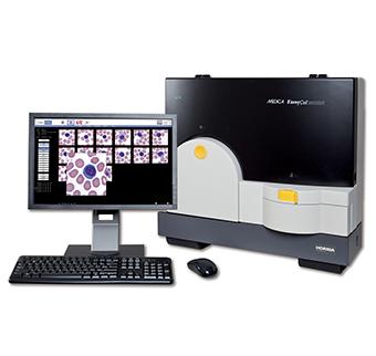
Product Description
The EasyCell locates and displays images of white cells, red cells and platelets. The typical scan time is approximately 4.15 minutes per slide for WBC only and 4.45 minutes per slide for WBC, RBC and Platelets. The Easy Cell uses sophisticated optical pattern recognition software to automatically locate and pre-classify normal white cells, smudge cells, NRBC’s and variant lymphs. These cells are then displayed and grouped by cell type for the technologist to confirm or reclassify the cells into the appropriate category.
The abnormal white cells are placed in the “Other” category the technologist can manually classify them. The pre-classification function reduces the operator’s time for classification. The EasyCell enables the operator to complete a WBC differential, RBC morphology evaluation, and platelet estimate using those images. The slides are introduced via the 30 slide carousel or STAT position. Special features available are the Competency review license used for training technologists and the Remote software license available for viewing the images remotely throughout the laboratory or hospital.
Features & Benefits
Improves productivity
The EasyCell assistant improves productivity by automatically locating 100 or 200 white cells on a blood smear and pre-classifying them on a display, grouped by cell type. The analyzer also displays images of red cells and platelets for performance of red cell morphology and platelet estimate.
Saves time
The EasyCell assistant lowers costs by dramatically reducing the technologist time required to perform manual differentials. EasyCell is an “assistant” because it suggests the correct classification of normal cells. It allows the technologist to quickly review the suggested classifications and then to devote time to the abnormal
Enables walk-away operation
Slides are loaded into the 30-position carousel or placed in the EasyCell Stat position for immediate analysis. Wright, Wright-Giemsa, or May-GrünwaldGiemsa stains may be used. The technologist walks away while slides are being processed.
Assists with accurate final classification
White cells are located and pre-classified for review on the display, grouped by cell type. Immature, abnormal and unrecognized cells are displayed separately. The technologist can easily compare white cell images which helps in final classification of cells. Red cell morphology images are reviewed and the technologist notes any abnormalities on the user interface. Platelet estimate and morphology can also be recorded.
Automates data access
Rapidly access slide files -recall previous patient’s cell images to track patient status. • View or print differential reports – add cell images and custom comments. • 1-D and 2-D bar coding, or label imaging for sample ID. • Store up to 10,000 slides on-board. • LIS connectivity — CLSI standards. • Archive data by saving a copy on a DVD, Thumb Drive, or External Hard Drive.
Assures accurate performance
EasyCell has built-in quality control software that verifies that nucleated cells are located and presented to the technologist for review. EasyCell quality control, run routinely, serves as a check for acceptable slide preparation and system hardware operation. It can also be run to verify performance when changes are made in stains or staining procedures.
Assists with teaching
Detailed cell images can easily be simultaneously viewed by instructor and student. Returning to cells of interest is easy.
Makes training easy
A simple user interface monitors and controls operation using only 5 screens: • Data Acquisition • Review List • White Blood Cells • Red Blood Morphology/Platelets • Quality Control Reduces eye strain and muscle fatigue. Automatic white cell location, pre-classification, and display of cells on a large digital screen makes it easy to see details.
